A Mechanical Model Involving the Spinal Dura Mater and the Suboccipital Muscles
For more than half a century respected authors, clinicians, and journals have claimed that various problems of the cervical spine can cause headaches. As an example, in 1958, Beverly Hills neurosurgeon Emil Seletz, MD, authors an article titled Headache of Extracranial Origin, published in the journal California Medicine (1). In this article, Dr. Seletz indicates that the cervical spine is an important cause of headache. He lists these potential origins for headache as:
1) Irritation of the C2 nerve root, which communicates with the trigeminal nerve to produce headache. C2 nerve root irritation occurs as a consequence of these mechanisms:
- Cervical trauma causing a “tractional injury” to the C2 nerve root because its exit is between C1 and C2, the point of “greatest rotation of the head on the neck.”
- Post-traumatic adhesions between the ligaments and the dural sleeve chronically irritating the C2 nerve root.
- Any cervical spine injury that may injure/irritate the delicate filaments of the spinal accessory nerves which originate from all points of the cervical spinal cord. This results in spasm of the trapezius muscles. Spasm of the trapezius muscle exerts traction on the greater occipital nerve at the point of where the nerve pierces the tendon of the trapezius muscle, resulting in occipital pain and headache.
2) Osteophytosis, swelling or adhesions arising from the uncinate process can irritate or compromise the vertebral artery or it’s sympathetic innervation. This has been a proposed mechanism of headache since 1925-1928, and is known as the syndrome of Barre-Lieou.
“Cervicogenic Headache” was officially recognized in 1983 (2). Most models of etiology for cervicogenic headache have involved the trigeminocervical nucleus and the convergence of the afferents of the trigeminal and upper cervical nerves:
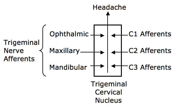
Australian clinical anatomist and physician Nikolai Bogduk, MD, PhD, champion this model of cervicogenic headache. In his 1995 article titled Anatomy and Physiology of Headache, Dr. Bogduk states (3):
“All headaches have a common anatomy and physiology. All headaches are mediated by the trigeminocervical nucleus, and are initiated by noxious stimulation of the endings of the nerves that synapse on this nucleus, by irritation of the nerves themselves, or by disinhibition of the nucleus.”
The trigeminocervical nucleus is “defined by its afferent fibers.” As its name implies, the primary afferent fibers into the trigeminocervical nucleus are from the trigeminal and upper cervical spine (C1-C3) sensory fields.
A New and Different Model for Cervical Headache
A careful review of Dr. Bogduk’s article (3) indicates that the spinal dura mater is innervated with nociceptors that arise from the upper cervical spine nerve roots. This innervation allows the spinal dura to be a generator of pain, primarily headache, as these afferents would synapse in the trigeminocervical nucleus.
Biomechanically, it is understood that when one extends their head or cervical spine, dural enfolding is produced. Rene Cailliet, MD, states (4):
“On extension the dura folds as in accordion pleats.”
[As of this writing (2012), Dr. Cailliet is still living at age 95].
White and Panjabi’s Clinical Biomechanics of the Spine (5) defines the pia mater as a “vascular membrane covering the [spinal] cord.” They also note that the pia mater is physically connected to the dura mater through the paired dentate ligaments:
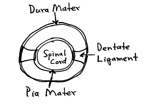
Consequently, cervical spine extension, especially abrupt extension, has the potential to irritate, compromise, injure, or inflame the spinal dura mater, the attached pia mater, and the spinal cord. Therefore, a protective mechanism whereby dural tension could be adapted to changes in head/neck positions is desirable.
••••
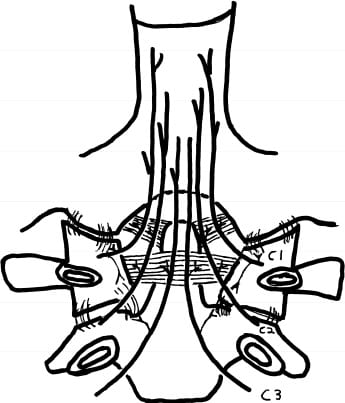
In 1992, French researchers investigated the detailed anatomy of the Atlanto-Occipital and Atlanto-Axial membranes (6). These researchers made two important discoveries:
- The fascia from the Rectus Capitis Posterior Minor muscle blends with the connective tissues of the Occipital-Atlanto membrane and they attach to the spinal dura mater.
- The fascia from the both the Rectus Capitis Posterior Major and Obliquus Capitis Inferior muscles blends with the connective tissues of the Atlanto-Axial membrane and they also attach to the spinal dura mater.
In summary, and important for this discussion, these researchers found that three of the four suboccipital muscles have fascial/connective tissue attachments to the spinal dura mater: Rectus Capitis Posterior Minor, Rectus Capitis Posterior Major, and Obliquus Capitis Inferior.

* Rectus Capitis Posterior Minor into the Occipital-Atlanto Space
** Rectus Capitis Posterior Major into the Atlanto-Axial Space
** Obliquus Capitis Inferior into the Atlanto-Axial Space
••••
In 1995, dentist Gary Hack, DDS, from the Baltimore College of Dental Surgery at the University of Maryland, published a pioneering study in the journal Spine titled (7):
Anatomic Relation Between the Rectus Capitis
Posterior Minor and the Dura Matter
In this study, Dr. Hack and colleagues carefully and meticulously dissected 11 human cadavers to investigate if there is a connection between the Rectus Capitis Posterior Minor muscle and the spinal dura mater. They note that the Rectus Capitis Posterior Minor muscle arises from the posterior arch of the atlas (C1) and inserts at the inferior nuchal line of the occiput. Deep to the Rectus Capitis Posterior Minor muscle is the Posterior Occipital-Atlanto Membrane, which is “intimately attached to the underlying spinal dura.”
ANATOMICAL FINDINGS:
Dense connective tissue attached the Rectus Capitis Posterior Minor muscle to the Posterior Occipital-Atlanto Membrane at the occipital-atlanto junction. The connective tissue bridge was arranged perpendicular to the dura. They state:
“A [dense] connective tissue bridge between the Rectus Capitis Posterior Minor muscle and the dorsal spinal dura at the atlanto-occipital junction was observed in every specimen.”
BIOMECHANICAL DISCUSSION:
Dr. Hack and colleagues found that head and neck extension of all cadavers in this study produced infolding of the Posterior Atlanto-Occipital Membrane/dura complex. When the head and neck are extended, the spinal dura folds inward toward the spinal cord. This enfolding “appeared to be resisted by this connective tissue bridge.” They state:
“The connective tissue bridge may help resist dural infolding during head and neck extension.”
These authors note that upper cervical spinal trauma causes atrophy of the Rectus Capitis Posterior Minor muscle. Such atrophy could compromise the resistance of dura infolding that occurs during head/neck extension. Thus, the dura could be irritated/injured/inflamed.
Immediately following this article was an attached Point of View by Scott Haldeman, DC, MD, PhD. Dr. Haldeman notes that injury or pathology affecting the cervical spine can cause headaches. The dura mater is a pain sensitive structure, capable of generating cervical headaches. Suboccipital muscle problems can influence the pain sensitive dura matter to generate headache. Massage, manipulation, and biofeedback directed to the cervical spine may be valuable in treating this headache mechanism. This is in agreement with Dr. Nikolai Bogduk, above (3).
••••
In 1997, HP Rutten and colleagues from the Department of Neurology, Deventer, The Netherlands, add to the discussion of Dr. Hack’s anatomical findings. Published in the journal Spine (8), Rutten and colleagues performed a layer-by-layer dissection of 7 human cadavers in an attempt to verify the upper cervical spine findings of Hack and colleagues. They state:
“The relation described by Hack et al between posterior atlanto-occipital membrane spinal dura complex and Rectus Capitis Posterior Minor is confirmed.”
Rutten et al suggest that the Rectus Capitis Posterior Minor muscle “monitors” stress on the dura mater. They further state:
“The Rectus Capitis Posterior Minor may function as a mechanoreceptive element, because it contains a great number of muscle spindles.”
“We hypothesize that the mechanism that helps to resist dural infolding and/or the proprioceptive registration fails in patients with whiplash-like symptoms.”
••••
In 1999, ME Alix and DK Bates, from Logan College of Chiropractic, reviewed the literature pertaining to the neurophysiological basis and anatomic relationship between the dura mater and the Rectus Capitis Posterior Minor muscle and the etiology of cervicogenic headache (9). They searched Medline and the Index to Chiropractic Literature. The title of their study is:
A proposed etiology of cervicogenic headache: the neurophysiologic basis and anatomic relationship between the dura mater and the rectus posterior capitis minor muscle
These authors note:
“Connective tissue bridges were noted at the atlanto-occipital junction between the rectus capitis posterior minor muscle and the dorsal spinal dura.
The perpendicular arrangement of these fibers appears to restrict dural movement toward the spinal cord.
Anatomic structures innervated by cervical spinal nerves C1-C3 have the potential to cause headache pain. Included are the joint complexes of the upper 3 cervical segments, the dura mater, and spinal cord.
A sizeable body of clinical studies note the effect of manipulation on headache. These results support its effectiveness.
The dura-muscular, dura-ligamentous connections in the upper cervical spine and occipital areas may provide anatomic and physiologic answers to the cause of cervicogenic headache.
This proposal would further explain manipulation’s efficacy in the treatment of cervicogenic headache.”
••••
In 2004, dentist Gary Hack and colleague report a case in which a patient experienced relief from chronic headache after surgical separation of the connective tissue bridge of the dura mater from the suboccipital musculature. Their case report was published in the journal Headache (10). This study would suggest that chronic suboccipital muscle tension could cause dysfunction, irritation, and/or irritation to the spinal dura mater, resulting is chronic headache. These authors state:
“The presence of a connective tissue bridge, attaching suboccipital muscles to the dura mater, is now recognized as a feature of normal human anatomy.”
••••
In 2005, Lance Nash and colleagues from the Department of Anatomy and Structural Biology and the School of Physiotherapy, University of Otago, New Zealand, published a study in the journal Spine titled (11):
Configuration of the Connective Tissue in the
Posterior Atlanto-Occipital Interspace
The objective of their study was to define the relationship between rectus capitis posterior minor, the posterior atlanto-occipital membrane, and the spinal dura in the posterior atlanto-occipital interspace. In their study the authors carefully dissected 22 human cadavers. They state:
“It has been speculated that connections between the dura and muscles and/or ligaments in the posterior atlanto-occipital interspace may transmit forces from the cervical spine joint complexes to the pain-sensitive dura, generating cervicogenic headaches.”
“Recently, the spinal dural connection of the rectus capitis posterior minor muscle and the nuchal ligament in the posterior atlanto-occipital interspace has been reported extensively.”
“The posterior atlanto-occipital membrane, extending between the posterior arch of the atlas (C1) and the occiput, has been observed to tether the spinal dura by numerous connective tissue fibers creating a posterior atlanto-occipital membrane-spinal dura complex.”
“The tendon of the rectus capitis posterior minor directly attaches to the spinal dura via the posterior atlanto-occipital interspace.”
“The present study demonstrates, for the first time, that tendinous fibers of the rectus capitis posterior minor muscle are continuous with the spinal dura.”
“A direct continuity of the rectus capitis posterior minor muscle to the spinal dura should have a great impact on strengthening the dura and preventing dural enfolding. Such an impact becomes particularly important during extension of the head and neck because, when the rectus capitis posterior minor muscle extends the cranio-cervical junction, a small portion of its muscular fibers simultaneously contract to pull the spinal dura posteriorly, preventing dural enfolding.”
“Microstrain and trauma in the rectus capitis posterior minor muscle and its tendon may cause a clonus condition of the muscle and stimulate the pain-sensitive dura, generating a cervicogenic headache.”
“The morphologic features of the rectus capitis posterior minor tendon and fascia indicate that they may have an important role in the maintenance of the posterior cranio-cervical stability and the prevention of the dural enfolding and are of anatomic relevance in the debate regarding the etiology of cervicogenic headaches.”
These authors found that the tendon of the rectus capitis posterior minor muscle fuses with the spinal dura via the posterior atlanto-occipital interspace. This study clearly demonstrates that the rectus capitis posterior minor tendon fibers are directly continuous with the spinal dura via the posterior atlanto-occipital interspace and become a part of the spinal dura.
••••
Most recently, in 2011, chiropractor Frank Scali, DC, and colleagues form the School of Medicine, St. George’s University, Grenada, West Indies, published a study in the journal Spine titled (12):
Anatomical Connection Between the Rectus Capitis
Posterior Major and the Dura Mater
Dr. Scali and colleagues note that chronic headaches in a significant number of patients are of cervical origin. Therefore, they did meticulous investigations bilaterally of 3 suboccipital muscles on 13 human cadavers:
Rectus Capitis Posterior Major (C2 spinous process to the occiput)
Rectus Capitis Posterior Minor (posterior arch C1 to the occiput)
Obliquus Capitis Inferior (C2 spinous process to the transverse process of C1)
They found:
- In 86% (11/13), the fibers from the Rectus Capitis Posterior Major entered the space between C1 and C2 and firmly attached to the dura.
- In 100% the fibers from the Rectus Capitis Posterior Minor entered the space between the occiput and C1 and firmly attached to the dura.
- In 86% (11/13) the fibers from the Obliquus Capitis Inferior entered the space (atlantoaxial interspace) between C1 and C2 and firmly attached to the dura.
Additionally, manual traction of the Rectus Capitis Posterior Major caused dural movement from the spinal root level of C2 to the spinal root level of T1.
Dr. Scali and colleagues note that mechanical stress applied to the dura mater during neurosurgical procedures results in cephalgia. Dural tension results in many clinical manifestations, especially headache. These authors suggest that proper function of the suboccipital muscles is required to prevent abnormal dural tension from occurring during various upper cervical spine motions, especially occiput-C1 flexion/extension and C1-C2 rotation.
••••
SUMMARY POINTS:
The 4 suboccipital muscles are innervated by the posterior primary rami of the C1 nerve root.
Three of the suboccipital muscles are directly and firmly attached to the spinal dura mater:
Rectus Capitis Posterior Major (C2 spinous process to the occiput)
Rectus Capitis Posterior Minor (posterior arch C1 to the occiput)
Obliquus Capitis Inferior (C2 spinous process to the transverse process of C1)
The apparent function of the attachment of the suboccipital muscles is to prevent the dura mater from being mechanically irritated, injured or inflamed during spinal motions.
The spinal dura mater is innervated with pain afferents from the upper cervical spine nerve roots.
Upper cervical spinal nerve root nociceptors synapse in the trigeminocervical nucleus, and are therefore capable of initiating headache.
Mechanical dysfunctions of the upper cervical spine may compromise the ability of the suboccipital muscles to protect the dura mater from motion related stress, irritation, injury, and inflammation.
Whiplash extension injuries occur quickly, so that the suboccipital muscles do not have enough time to contract and pull the spinal dura to safety, resulting in injury and headache.
In chronic whiplash patients, injured suboccipital muscles may undergo atrophy and fatty infiltration, further compromising the ability of these muscles to protect the dura mater from irritation and inflammation during routine motions, resulting in headache.
Chronic upper neck postural stress and distortions that invoke contraction of the suboccipital muscles may cause chronic stress on the spinal dura mater, resulting in headache.
Mechanical dysfunctions of the upper cervical spine may also irritate/inflame the spinal cord and its blood supply because the dura mater is attached to the vascular pia mater that surrounds the spinal cord.
There is biological plausibility for upper cervical spinal manipulation, occiput-atlas-axis chiropractic alignment, postural improvement, and suboccipital muscle myotherapy to be utilized in the cervicogenic headache patient.
REFERENCES:
- Seletz E; Headache of Extracranial Origin; California Medicine; November 1958, Vol. 89, No. 5, pp. 314-317.
- Sjaastad O; “Cervicogenic Headache” An Hypothesis; Cephalagia; December 1983; 3(4):249-256.
- Bogduk N; Anatomy and Physiology of Headache; Biomedicine and Pharmacotherapy; 1995, Vol. 49, No. 10, 435-445.
- Cailliet R; Neck and Arm Pain, Edition 2; FA Davis Company; Philadelphia; 1982.
- White AA, Panjabi M; Clinical Biomechanics of the Spine; Second edition; Lippincott; Phildelphia; 1990.
- Kahn JL, Sick H, Koritke JG; [The posterior intervertebral spaces of the craniovertebral joint]; Acta Anat (Basel); 1992;Vol. 144; No. 1; pp. 65-70.
- Hack G, Koritzer R, Robinson W, Hallgren R, Greenman P; Anatomic Relation Between the Rectus Capitis Posterior Minor and the Dura Matter; Spine; December 1, 1995; Vol. 20; No. 23; pp. 2484-2486.
- Rutten HP, Szpak K, van Mameren H, Ten Holter J, deJong J; Letters: comment on Anatomic Relation Between the Rectus Capitis Posterior Minor and the Dura Matter; Spine, April 15, 1997; Vol. 22; No. 8; pp. 924-926.
- Alix ME, Bates DK; A proposed etiology of cervicogenic headache: the neurophysiologic basis and anatomic relationship between the dura mater and the rectus posterior capitis minor muscle; Journal of Manipulative and Physiological Therapeutics; October 1999. Vol. 22; No. 8; pp. 534-539.
- Hack GD, Hallgren RC; Chronic headache relief after section of suboccipital muscle dural connections: a case report; Headache; January 2004;44(1):84-9.
- Nash L, Nicholson H, Lee ASJ, Johnson GM, Zhang M; Configuration of the Connective Tissue in the Posterior Atlanto-Occipital Interspace; Spine; Volume 30(12) June 15, 2005 pp. 1359-1366
- Scal F, Marsili ES, Pontell ME; Anatomical Connection Between the Rectus Capitis Posterior Major and the Dura Mater; Spine; December 1, 2011; Vol. 36; No. 25, pp. E1612–E1614.